- Veterinary View Box
- Posts
- Where do the blood goes if there is no caudal vena cava?
Where do the blood goes if there is no caudal vena cava?
JSAP 2009
T Schwarz 1, F Rossi, J D Wray, B Ablad, M W Beal, J Kinns, G S Seiler, R Dennis, J F McConnell, M Costello
Background
Segmental caudal vena cava (CVC) aplasia is a rare congenital vascular anomaly in dogs, characterized by interruption or absence of a segment of the CVC. The missing postrenal caval segment results in venous blood being shunted to an azygos vein. While previous case reports have described this anomaly, there is limited data on computed tomographic angiography (CTA) and magnetic resonance angiography (MRA) features. This study aimed to describe the imaging characteristics of CVC aplasia and its associated vascular anomalies in affected dogs.
Methods
Study Design: Retrospective review of CT and MRI records from eight veterinary referral institutions between 1999 and 2007.
Inclusion Criteria:
-A diagnostic-quality abdominal CTA or MRA study.
-Supportive clinical and follow-up data.
Data Collection:
-Signalment, clinical history, and imaging findings.
-CTA and MRA studies were evaluated for:
-Size, shape, location, and course of abdominal veins.
-Presence of shunt vessels, thrombosis, and abnormal drainage patterns.
Results
Population:
-10 dogs met the inclusion criteria.
-Mean age: 2.6 years (range: 3 months to 8 years).
-70% of affected dogs were female.
Imaging Findings:
-All dogs had a missing postrenal CVC segment, with venous drainage redirected to the right azygos vein (8/10) or left azygos vein (2/10).
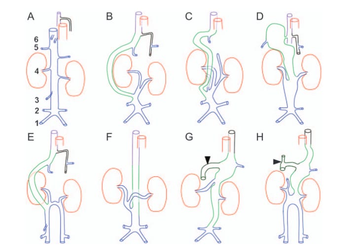
-Seven distinct angiographic patterns of cavo-azygos shunting were identified.
-Aneurysmal dilation of the shunt vessel was observed in 40% of cases, with thrombus formation in one dog.
-Two cases had additional porto-azygos shunts, where portal blood drained into the azygos vein instead of the liver.
-One dog exhibited multiple vascular anomalies, including an intrahepatic portocaval shunt and complete azygos continuation of the CVC.
Clinical Associations:
-Most dogs were asymptomatic, with the anomaly detected incidentally during imaging for unrelated conditions.
-Two dogs presented with episodic collapse, possibly related to impaired venous return through an aneurysmal, collapsing shunt vessel.
-One dog developed thrombosis in the shunt vessel, highlighting the potential for vascular complications.
Limitations
Small sample size (n = 10), limiting generalizability.
Retrospective study design, dependent on the availability and quality of archived imaging data.
Lack of histopathology or genetic analysis, preventing a deeper investigation into the developmental basis of CVC aplasia.
Conclusions
Segmental CVC aplasia results in postrenal venous blood shunting to an azygos vein, with multiple anatomic variations observed on CTA and MRA. Although often an incidental finding, some cases may present with thrombosis, aneurysmal dilation, or collapse-related venous insufficiency. CTA and MRA provide excellent visualization of complex vascular anomalies, making them essential for preoperative planning in dogs with concurrent portosystemic shunts or thromboembolic disease.

Schematic drawings of abdominal caval anatomy and seven types of segmental aplasia of thecaudal vena cava (CVC) and shunting viewed ventrally (left side on image right). Blue, CVC andtributary veins (1, common iliac; 2, deep circumflex iliac; 3, right testicular/ovarian; 4, renal; 5,phrenicoabdominal and 6, hepatic veins); brown, kidneys; red, aorta; magenta, right azygos vein;black, hemiazygos/left azygos vein; light green, cavo-azygos shunt vessel; dark green, portal shuntvessel (arrowhead) connecting to cavo-azygos shunt. Details of portal and hepatic caval veinanatomy/anomaly are not depicted in this drawing. (A) Normal anatomy. (B) Type 1 (case 1), rightlateral cavo-right-azygos shunt. (C) Type 2 (case 2), right medial cavo-right-azygos shunt and smallblind ending CVC cranial to left kidney. (D) Type 3 (case 3; cases 8, 9 and 10 were similar, exceptdetails of tributary veins), large aneurysmal right medial cavo-right-azygos shunt with an isthmianconnection to the azygos vein. (E) Type 4 (case 4), split CVC and right medioventral cavo-right-azygos shunt. (F) Type 5 (case 5), dorsal cavo-right-azygos shunt. (G) Type 6 (case 6), aneurysmalcavo-left-azygos shunt with connecting completely shunting portal vein. (H) Type 7 (case 7), splitCVC, aneurysmal cavo-left-azygos shunt and connecting portal vein shunt vessel
How did we do? |
Disclaimer: The summary generated in this email was created by an AI large language model. Therefore errors may occur. Reading the article is the best way to understand the scholarly work. The figure presented here remains the property of the publisher or author and subject to the applicable copyright agreement. It is reproduced here as an educational work. If you have any questions or concerns about the work presented here, reply to this email.