- Veterinary View Box
- Posts
- You can see those thrombus on CT too
You can see those thrombus on CT too
Veterinary Radiology & Ultrasound, 2018.
Kyle P. Vititoe, Ryan C. Fries, Stephen Joslyn, Laura E. Selmic, Mark Howes, Jordan P. Vitt, Robert T. O'Brien.
Background
Arterial thromboembolism is a severe complication of feline cardiac disease, often originating from thrombi in the left atrium. Echocardiography is the standard method for diagnosing intra-cardiac thrombi, but computed tomographic (CT) angiography is gaining use in humans for detecting left atrial thrombi. This study aimed to evaluate the feasibility of CT angiography in awake cats with cardiac disease, compare its accuracy to echocardiography in detecting thrombi, and assess its ability to identify prognostic factors and comorbidities.
Methods
A prospective pilot study was conducted on 14 cats with heart disease, seven with confirmed thrombi and seven with spontaneous echocardiographic contrast. Transthoracic echocardiography served as the reference standard. All cats underwent CT angiography using a microdose contrast protocol while awake or lightly sedated. Two imaging techniques were assessed: continuous CT angiography and dynamic (cine-loop) CT imaging. CT scans were reviewed by two radiologists and two cardiologists blinded to clinical data. Sensitivity, specificity, and interobserver agreement were analyzed.
Results
CT angiography detected six of seven thrombi as filling defects, primarily in the left auricle (n = 6) and right heart (n = 1). Continuous CT had a sensitivity of 71.4% and specificity of 71.4%, while dynamic CT had a lower sensitivity (57.1%) but higher specificity (85.7%). Interobserver agreement was fair to moderate (κ = 0.38–0.44). CT angiography also identified prognostic factors, including left atrial enlargement, congestive heart failure, and arterial thromboembolism. Additional comorbidities detected included suspected idiopathic pulmonary fibrosis and asthma.
Limitations
The small sample size limited statistical power. Histopathological confirmation of thrombi was available for only three cases. Motion artifacts and partial volume effects reduced the accuracy of thrombus detection, particularly in small, mobile, or incompletely occlusive thrombi. Reviewers had limited prior experience evaluating feline left auricles on CT, potentially affecting sensitivity.
Conclusions
CT angiography is a viable method for detecting intra-cardiac thrombi, identifying cardiac enlargement, and diagnosing congestive heart failure in awake or lightly sedated cats. Continuous-phase CT is more sensitive, while dynamic imaging offers higher specificity. Though not a replacement for echocardiography, CT angiography provides additional diagnostic and prognostic information, particularly in cats with respiratory distress or when echocardiography is unavailable.

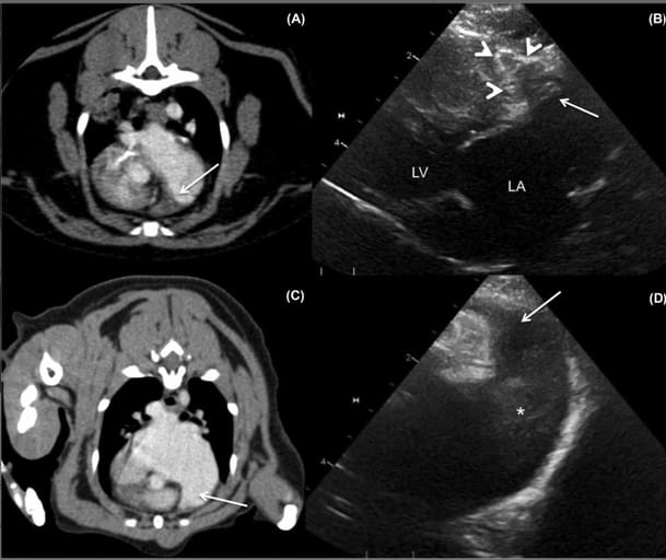
A, Transverse computed tomographic angiography of a patient with a dilated left atrium and left auricle and a large filling defect (presumed thrombus) (white arrow) occupying most of the left auricle. B, Oblique left apical parasternal long-axis echocardiographic image of the left auricle with corresponding thrombus that is similar in appearance (conforming to and occupying most of the left auricle). The white arrowheads outline the external margins of the left auricle. LA, left atrium; LV, left ventricle. The second set of images (C,D) is of a patient with severe left atrial enlargement and only spontaneous echo contrast identified on echo. C, Transverse thoracic computed tomographic angiography and oblique left apical parasternal long-axis view of the left auricle (D) with severely dilated left atrium/left auricle with no filling defects (thrombi) noted within the left auricle (white arrow). The asterisk highlights the spontaneous echo contrast within the left atrium. The right of the patient is on the left in the CT images and displayed in soft tissue window (window length, 40; window width, 400)
How did we do? |
Disclaimer: The summary generated in this email was created by an AI large language model. Therefore errors may occur. Reading the article is the best way to understand the scholarly work. The figure presented here remains the property of the publisher or author and subject to the applicable copyright agreement. It is reproduced here as an educational work. If you have any questions or concerns about the work presented here, reply to this email.